

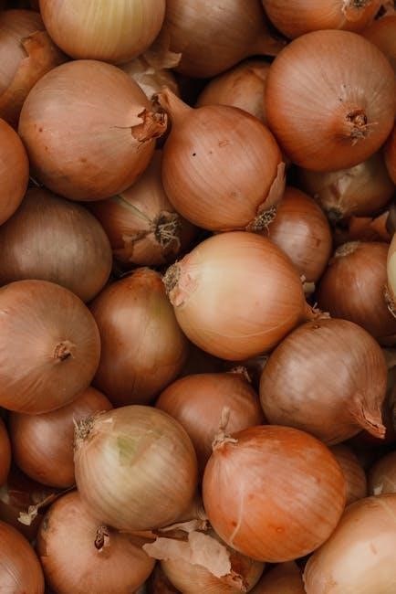
Mitosis is a cell division process producing identical daughter cells. Observing onion root tips helps study mitosis due to their actively dividing meristematic cells, making them ideal for educational labs.
1.1 Definition and Importance of Mitosis
Mitosis is a type of cell division that results in two genetically identical daughter cells. It is crucial for growth, tissue repair, and asexual reproduction. In the onion root tip experiment, mitosis is studied to observe its stages under a microscope. The process helps understand cellular behavior, chromosome distribution, and the cell cycle. Observing mitosis in onion root tips is ideal due to their actively dividing meristematic cells, providing a clear view of each phase. This experiment aids in teaching cell biology concepts and the importance of mitosis in plant growth and development. It also highlights the role of mitosis in maintaining genetic continuity across generations.
1.2 Brief Overview of the Onion Root Tip Experiment
The onion root tip experiment is a classic biology lab used to study mitosis. It involves observing the stages of cell division in the meristematic region of onion roots. The process includes fixing the root tips in a solution to preserve cells, staining them with a dye like toluidine blue to enhance visibility, and preparing slides for microscopy. This experiment allows students to identify and count cells in different phases of mitosis. It demonstrates the cell cycle’s progression and the relative duration of each phase. The experiment is simple, cost-effective, and provides hands-on experience with cellular biology concepts. It is widely used in educational settings to teach mitosis and its significance in plant growth.

The Cell Cycle in Onion Root Tips
The cell cycle in onion root tips is a continuous process of growth, DNA replication, and cell division, enabling root elongation and development.

2.1 Phases of the Cell Cycle
The cell cycle consists of four main phases: G1 (gap 1), S (synthesis), G2 (gap 2), and M (mitosis). In onion root tips, these phases occur sequentially. G1 involves cell growth and preparation for DNA replication, while the S phase is dedicated to DNA synthesis. G2 allows the cell to finalize preparations for mitosis. The M phase includes mitosis and cytokinesis, where the cell divides into two daughter cells. These phases ensure controlled growth and development in root tips, making them ideal for studying mitosis due to their high division rate and synchronized cell cycles.
2.2 Duration of the Cell Cycle in Onion Root Cells
The duration of the cell cycle in onion root cells varies but typically ranges from 12 to 36 hours, depending on environmental conditions. The G1 phase is the longest, followed by the S phase, G2 phase, and finally the M phase. Mitosis (M phase) is the shortest, lasting about 1-2 hours. The cell cycle in root tips is influenced by factors such as temperature, nutrient availability, and growth hormones. Understanding the duration helps in timing the experiment correctly, ensuring cells are in different stages of division when observed. This variability highlights the dynamic nature of cell growth in root tips.
2.3 Why Root Tips Are Ideal for Observing Mitosis
Root tips are ideal for observing mitosis due to their high rate of cell division, making it easier to capture cells in various stages of mitosis. The meristematic region at the root tip contains undifferentiated cells that divide rapidly, increasing the likelihood of observing multiple stages under a microscope. Additionally, root tips are easy to obtain, handle, and prepare for observation. They can be fixed, stained, and sectioned without complex procedures, making them suitable for educational labs. The large size and clear cytological features of root tip cells enhance visibility, allowing students to distinguish between different phases of mitosis effectively. This accessibility and visibility make root tips a practical choice for studying the process of mitosis.

Preparation for the Experiment
Preparation involves fixing root tips to preserve cells, staining to enhance visibility under a microscope, and sectioning or squashing to prepare slides for observation.
3.1 Materials Required
To conduct the onion root tip mitosis experiment, several materials are essential. These include onion bulbs, a compound microscope, glass slides, cover slips, forceps, a measuring cylinder, a beaker, Carnoy’s solution (ethanol and hydrochloric acid mixture), staining dye (e.g., orcein or methylene blue), distilled water, paper towels, and a clean workspace. Additional items like a sharp razor or scalpel are needed for cutting root tips, while a microscope light source ensures clear visibility. These materials are critical for preparing, preserving, and observing the root tip cells to study mitosis effectively.
3.2 Step-by-Step Procedure for Preparing the Specimen
Begin by cutting 1-2 cm of the onion root tip using a sharp razor or scalpel. Immediately place the root tips in Carnoy’s solution (ethanol and hydrochloric acid mixture) for 4-24 hours to fix and preserve the cells. After fixation, rinse the root tips in distilled water to remove excess solution. Soak them in 0.1M HCl for 10-15 minutes to soften the tissue. Then, transfer the root tips to a staining solution (e.g., orcein or methylene blue) for 1-2 hours to enhance cell visibility. Finally, prepare the slide by placing the root tip on a slide, squashing it gently with a cover slip, and observing under a microscope.
3.3 Fixation and Staining of Root Tips
Fixation is done using Carnoy’s solution, a mixture of ethanol and hydrochloric acid, to preserve the cells and stop metabolic processes. This step ensures that the cells are fixed in their current phase of mitosis. After fixation, the root tips are rinsed in distilled water to remove excess solution. Staining is then performed using a dye like orcein or methylene blue, which binds to DNA and proteins, making the cell structures visible under a microscope. The staining step is critical for distinguishing cellular components, especially chromosomes, during mitosis. Proper fixation and staining are essential for clear observations and accurate data collection.
Observing Mitosis Under the Microscope
Under the microscope, mitotic cells are identified in the meristematic region. Cells in various stages of mitosis are located, and their structures are analyzed for study.
4.1 Setting Up the Microscope
To observe mitosis, the microscope must be properly set up. Begin by assembling the microscope and preparing the slide with the stained root tip. Place the slide on the stage and secure it with clips. Focus on the sample using low magnification (4x or 10x objective lens) to locate the meristematic region. Adjust the condenser and lighting for optimal clarity. Once focused, switch to high magnification (40x objective lens) for detailed observation. Ensure the stage is properly aligned, and the light intensity is adjusted for clear visibility. Proper setup is crucial for accurate cell identification and analysis during the experiment.
4;2 Identifying the Meristematic Region
The meristematic region, located near the tip of the onion root, is where active cell division occurs. To identify it, focus on the white, growing tip of the root under the microscope. This region contains small, densely packed cells with minimal vacuoles, making it ideal for observing mitosis. Use low magnification to locate the tip, then switch to high magnification for clearer detail. The cells in this area are smaller and more rectangular compared to differentiated cells, with visible nuclei. Proper identification ensures accurate observation of mitotic stages, as this region has the highest concentration of dividing cells.
4.3 Locating Cells in Different Stages of Mitosis
To locate cells in different stages of mitosis, systematically scan the meristematic region under high magnification. Begin by identifying cells with visible nuclei, as these are likely in interphase. Transition to cells showing chromatin condensation for prophase, followed by metaphase cells with chromosomes aligned at the center. Anaphase cells will display separated chromosomes, while telophase cells show nuclear envelope reformation. Cytokinesis, the final stage, is marked by cell wall formation. Since most cells are in interphase, patience is needed to find cells in later stages. Carefully examine multiple fields of view to capture diverse stages, ensuring accurate observations for analysis.

The Phases of Mitosis
Mitosis consists of six main stages: interphase, prophase, metaphase, anaphase, telophase, and cytokinesis. These stages ensure proper cell division and genetic material distribution to daughter cells.
5.1 Interphase
Interphase is the longest stage of mitosis, where the cell grows, replicates its DNA, and prepares for cell division. It consists of three sub-phases: G1 (growth), S (DNA synthesis), and G2 (preparation for mitosis). During this phase, the chromatin remains loosely packed, and no visible chromosomes are present. In onion root tip cells, interphase nuclei appear large and dark due to DNA condensation. This phase ensures that the cell is ready for the subsequent stages of mitosis by producing essential proteins and organelles. Observing interphase cells is crucial for understanding the cell cycle progression in the onion root tip experiment.
5.2 Prophase
Prophase is the first stage of mitosis, marking the beginning of visible chromosomal changes. During this phase, chromatin condenses into visible chromosomes, and the nucleolus disappears. The nuclear envelope begins to break down, releasing nuclear materials into the cytoplasm. Spindle fibers form, extending from the centrioles to attach to the centromeres of the chromosomes. These spindle fibers prepare the chromosomes for alignment during metaphase. In onion root tip cells, prophase is identifiable by the appearance of condensed chromosomes and the initial formation of the spindle apparatus. This stage is critical as it sets the stage for the precise distribution of chromosomes during mitosis.
5.3 Metaphase
Metaphase is the second stage of mitosis, characterized by the alignment of chromosomes at the cell’s equatorial plane. Spindle fibers attach to the centromeres of sister chromatids, ensuring proper alignment. This phase ensures that each daughter cell will receive an identical set of chromosomes. The metaphase plate forms, and chromosomes are poised for separation. In onion root tip cells, metaphase is easily identifiable by the aligned chromosomes and the visible spindle fibers. This stage is critical for ensuring genetic continuity and is brief, lasting only until the chromosomes are properly positioned. Observing metaphase cells under the microscope is straightforward due to their distinct alignment.
5.4 Anaphase
Anaphase is the third stage of mitosis, where sister chromatids are pulled apart by spindle fibers toward opposite poles of the cell. This separation ensures each daughter cell receives an identical set of chromosomes. The chromatids, now considered individual chromosomes, move at a constant speed during this brief phase. In onion root tip cells, anaphase is easily recognizable by the visible separation of chromosomes. This phase is critical for ensuring genetic continuity and occurs rapidly, lasting only a few minutes. Observing anaphase in root tips provides clear evidence of chromosomal segregation, a fundamental aspect of mitosis. This stage is shorter than interphase but equally vital for cell division.
5.5 Telophase
Telophase is the fourth stage of mitosis, marking the transition toward the completion of cell division. During this phase, the nuclear envelope reforms around each set of chromosomes, and the spindle fibers dissolve. The chromosomes, now at opposite poles of the cell, uncoil and revert to their less condensed chromatin state. Nucleoli also reappear, signifying the restoration of nuclear structure. In plant cells, such as those in onion root tips, the beginning of cytokinesis is evident as the cell plate forms, eventually dividing the cytoplasm. Telophase ensures the daughter cells inherit the necessary genetic material and organelles, preparing them for interphase. This phase is crucial for restoring cellular normalcy and completing mitosis.
5.6 Cytokinesis
Cytokinesis is the final stage of mitosis, where the cytoplasm divides, and the cell splits into two daughter cells. In plant cells, such as those in onion root tips, a cell plate forms at the equator of the cell, gradually expanding to separate the cytoplasm. This process begins during telophase and completes shortly after. Vesicles from the Golgi apparatus fuse to form the cell plate, which develops into a new cell wall. The result is two genetically identical daughter cells, each with a complete set of organelles and chromosomes. Cytokinesis ensures the proper distribution of cellular components, completing the division process observed in the meristematic region of the root tip.
Data Collection and Analysis
Data collection involves counting cells in each mitotic phase under the microscope, recording observations, and calculating percentages. This step is crucial for understanding mitotic activity patterns.
6.1 Counting Cells in Each Phase
Counting cells in each phase involves systematically scanning the meristematic region under the microscope. A tally system is used to record the number of cells in interphase, prophase, metaphase, anaphase, telophase, and cytokinesis. Multiple fields of view are observed to ensure accuracy and representation. Cells are identified based on chromosomal appearance and arrangement. This step requires careful observation to avoid miscounting. The total number of cells counted typically ranges between 300-500 for reliable data. The process helps determine the relative duration of each phase. Consistency is key to minimize errors. This data forms the basis for calculating percentages in subsequent steps.
6.2 Calculating the Percentage of Cells in Each Phase
Calculating the percentage of cells in each phase involves dividing the number of cells in a specific phase by the total number of cells counted, then multiplying by 100. This provides a proportional representation of cells in each stage. For example, if 120 cells are in interphase out of 500 total cells, the percentage is (120/500) × 100 = 24%. This step helps visualize the distribution of cells across mitotic phases. The percentages are used to compare with the expected durations of each phase. Accurate calculations are crucial for reliable data analysis. This method ensures a clear understanding of cellular behavior during mitosis.
6.3 Determining the Longest Phase of Mitosis
Determining the longest phase of mitosis involves analyzing the percentage of cells in each phase. Since most cells are in interphase, it is the longest phase. By comparing the percentages, interphase is identified as the most time-consuming stage due to DNA replication and protein synthesis. Other phases, like prophase and metaphase, are shorter and more dynamic. This analysis aligns with the cell cycle’s duration, where interphase occupies about 90% of the cycle. Understanding the longest phase helps in grasping the cell cycle’s efficiency and the importance of preparation for cell division. This step is crucial for interpreting mitotic activity in the onion root tip experiment.
Results and Discussion
The results showed most cells in interphase, indicating its prolonged duration. This reflects the cell cycle’s focus on growth and preparation phases for cell division.
7.1 Sample Data Table and Observations
A sample data table from the experiment is shown below:
| Phase of Mitosis | Number of Cells Observed |
|---|---|
| Interphase | 150 |
| Prophase | 20 |
| Metaphase | 15 |
| Anaphase | 10 |
| Telophase | 12 |
| Cytokinesis | 8 |
Observations revealed that most cells were in interphase, while fewer cells were in the later stages of mitosis. This aligns with the fact that interphase is the longest phase. Additionally, distinguishing between prophase and metaphase was challenging due to similar chromatin structures. Anaphase and telophase cells were less frequent, reflecting their shorter durations. Cytokinesis was rarely observed, as it occurs simultaneously with telophase. These findings are consistent with the expected distribution of cells in the cell cycle.
7.2 Comparison with the Cell Cycle Duration

The experimental data aligns with the known durations of the cell cycle phases. Interphase, being the longest phase, comprised the majority of observed cells (150 out of 215), consistent with its 90-95% duration of the cycle. Prophase, metaphase, anaphase, and telophase were less frequent, reflecting their shorter durations. For example, metaphase and anaphase each lasted only a few minutes, which is why fewer cells were observed in these stages. Cytokinesis, occurring simultaneously with telophase, was rarely captured. This distribution confirms that cells spend most of their time in interphase, preparing for division. The findings support the concept that mitosis is a brief but critical phase of the cell cycle.
7.3 Significance of the Findings
The findings of the onion root tip experiment are significant as they provide direct observational evidence of mitosis in plant cells. This experiment helps students understand the cell cycle and the distribution of cells in different phases. By quantifying cells in each stage, the experiment demonstrates that interphase is the longest phase, while mitosis itself is relatively brief. This aligns with the biological concept that most of a cell’s life is spent in preparation for division. The experiment also highlights the importance of mitosis in plant growth, particularly in root development. Such studies are foundational for understanding cellular biology and have practical applications in genetics and agriculture.

Common Questions and Answers
Why are onion root tips used? They have meristematic cells that actively divide; What is the role of hydrochloric acid? It softens cell walls for staining. Challenges include identifying mitotic phases accurately due to their brief duration and similar appearances under a microscope.
8.1 Which Phase of Mitosis Lasts the Longest and Why?
The interphase lasts the longest in mitosis, accounting for 90-95% of the cell cycle duration. During interphase, the cell grows, replicates its DNA, and synthesizes proteins essential for division. This phase is longer because DNA replication and preparation for cell division are complex processes requiring significant time. In contrast, prophase, metaphase, anaphase, and telophase are shorter, as they involve rapid structural changes. Interphase ensures the cell is ready to divide, making it the most time-consuming phase. Observing this in onion root tips highlights the cell cycle’s efficiency and the importance of proper preparation for mitosis.
8.2 How Does the Microscope Magnification Affect Observations?
Microscope magnification significantly impacts the clarity and detail of observations in the onion root tip experiment. Higher magnification (e.g., 400x or 1000x) allows for better visualization of cellular structures and mitotic phases, such as chromatin condensation or chromosome alignment. However, excessive magnification can reduce the field of view, making it harder to locate the meristematic region. Lower magnification (e.g., 100x) provides a broader view, aiding in the initial identification of dividing cells. Balancing magnification is crucial for accurate observations, as it ensures both detail and context are maintained. Proper adjustment enhances the ability to distinguish between mitotic stages accurately.
8.3 Why Are Root Tips Ideal for Studying Mitosis?
Root tips are ideal for studying mitosis due to their high concentration of meristematic cells, which actively divide to produce new tissues. These cells are small, uniformly sized, and densely packed, making them easier to stain and observe under a microscope. The root tip’s organization into distinct zones, such as the apical meristem, allows for clear identification of cells in various stages of mitosis. Additionally, root tips are readily available, inexpensive, and easy to prepare, making them a practical choice for educational labs. Their continuous growth ensures a high mitotic index, increasing the likelihood of observing dividing cells in different phases.
Challenges and Limitations
Challenges include identifying mitotic phases due to their brief duration, small cell size, and overlapping stages. Incorrect preparation can distort results, and novice students may struggle distinguishing phases accurately.
9.1 Difficulty in Identifying Certain Phases
One major challenge is distinguishing between mitotic phases due to their similarity. Interphase and telophase are often confused as they share nuclear features. Prophase and metaphase may also appear alike because chromatin condensation can vary. Root tip cells are small, making detailed observation hard. Additionally, improper staining or preparation can obscure cellular structures, leading to misidentification. Novice students often struggle to accurately classify phases, especially anaphase, which is brief and less common. These factors highlight the need for careful sample preparation and skilled observation to ensure accurate phase identification during the experiment.
9.2 Potential Sources of Error
Errors in the onion root tip mitosis lab can arise from improper fixation or staining, leading to poor cell preservation or weak chromatin visibility. Using outdated or low-quality materials, such as dull microscopes or ineffective dyes, can reduce observation accuracy. Overcrowding of cells on slides makes it hard to distinguish individual cells. Additionally, human error during counting or phase identification can skew results. Inadequate training or rushing the procedure increases the likelihood of mistakes. Ensuring precise techniques, using fresh root tips, and maintaining optimal staining times are critical to minimizing errors and achieving reliable data for analysis.
9.3 Improving the Experiment
To enhance the onion root tip mitosis experiment, several improvements can be implemented. Using higher-quality microscopes with better resolution can improve cell visibility. Incorporating additional stains or fluorescent dyes could enhance chromatin detail. Providing more extensive training on slide preparation and cell identification reduces human error. Increasing the number of root tip samples and observing more cells ensures statistically significant data. Implementing a digital counting tool or software can minimize manual counting errors. Additionally, optimizing fixation and staining times, as well as using enzymatic methods to soften cell walls, can improve cell separation and observation clarity. These adjustments enhance accuracy and reliability in the experiment.
The onion root tip experiment successfully demonstrates mitosis stages, enhancing understanding of cellular division. It highlights the cell cycle’s importance and practical lab techniques for biology education.
10.1 Summary of Key Takeaways
The onion root tip experiment effectively demonstrates the stages of mitosis, providing insights into the cell cycle. Key observations include the identification of interphase, prophase, metaphase, anaphase, telophase, and cytokinesis. The experiment highlights the importance of proper specimen preparation, including fixation and staining, to visualize cellular structures clearly. Data analysis revealed that most cells are in interphase, while fewer cells are in later stages, reflecting the duration of each phase. This lab reinforces understanding of mitotic processes and their significance in growth and development. It also emphasizes the value of microscopy in biological studies and the practical application of statistical analysis in interpreting cellular data.
10.2 Practical Applications of the Experiment
This experiment offers valuable practical insights into cellular biology, particularly in education and research. It serves as a hands-on tool for teaching mitosis and the cell cycle, helping students grasp complex biological processes. The techniques learned, such as microscopy and specimen preparation, are foundational skills for future scientific studies. Additionally, understanding mitosis is crucial in fields like cancer research, agriculture, and genetics. The experiment also illustrates the importance of statistical analysis in biology, making it a comprehensive learning experience. These skills and concepts are essential for advancing knowledge in life sciences and developing practical applications in medicine and biotechnology.

10.3 Importance of Understanding Mitosis
Understanding mitosis is fundamental for comprehending cellular biology and its role in growth, development, and disease. Mitosis is essential for tissue repair, organism development, and asexual reproduction. Errors in mitosis can lead to uncontrolled cell division, often resulting in cancer. Studying mitosis helps in developing cancer treatments and understanding genetic disorders. It also aids in agricultural practices, such as plant breeding, to improve crop yields. Furthermore, mitosis is crucial for regenerative medicine and understanding how organisms heal. This knowledge provides insights into life processes and informs advances in medical and biological sciences, making it a cornerstone of modern biology education and research.